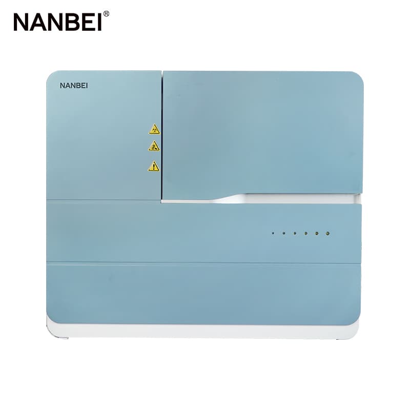
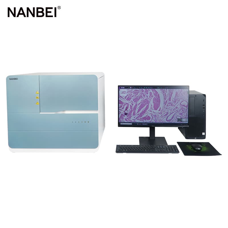
Pathology Slide Scanners
NANBEI Is Professional On Providing One-step Solution Of Laboratory Instruments And Equipment
Brand:NANBEI
Model:
Application:
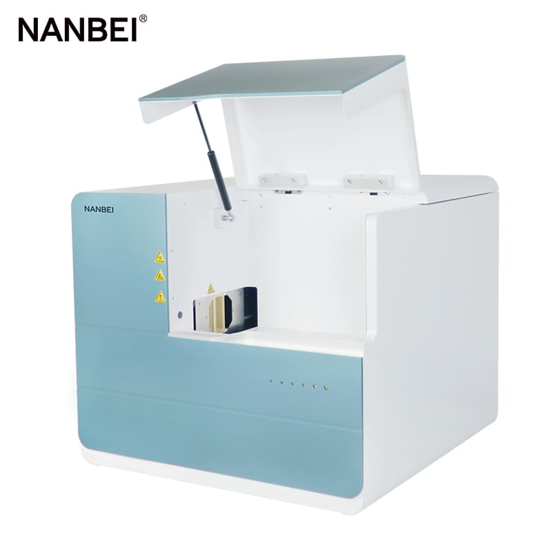
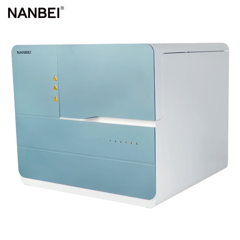
Laboratory Histology Pathological Tissue Slide Scanner
Products Description

Digital Benefits
Digital slicing is the rapid scanning of pathological slide specimens into high-resolution electronic pictures, that is, the process of rapid digitization of material slide specimens. Its essence is to achieve standardized, high-definition, and full-information image acquisition;
Convenience for slice reading—make the film reading free from the limitation of the microscope, mouse operation can quickly select any part of the slice for observation, without being limited by the field of view under the microscope, with complete structure, comprehensive field of view, high resolution, clear pictures, saturated colors, and undistorted images ;
Slice preservation—analyze and classify similar pathological slice digital images through analysis and statistical software, making search and management more convenient, efficient and convenient. It avoids the breakage and discoloration of traditional slices in the process of production and use. Some precious specimens can be stored for a long time after being converted into digital slices, and they are not easy to lose.
Scientific research analysis—to ensure the consistency of image quality and the accuracy of each analysis data;

Based on the usage habits of scientific research users, the software is adjusted, and the results are exported according to the requirements of scientific research articles and reports;
Slice preview information can be retrieved at any time, and panoramic result format conversion is supported, which facilitates the versatility of result analysis.
Provide a complete secondary development interface, which can integrate artificial intelligence identification schemes in the instrument software system to realize functions such as digital diagnosis, intelligent identification, and automatic result interpretation.

Specifications
Details
 | S-shaped track area scan |
Panorama storage compression is smallerHigh-speed, efficient and unattended Based on the extreme compression technology of the special database of artificial intelligence algorithm, the storage space and cost are greatly saved without affecting the image quality. |  |
 | Fast autofocusWide-range focal plane fitting, field curvature digital correction Intelligently select the focus position according to the tissue situation, and fit the focal plane of the entire tissue according to the selected focus value to ensure that the image of each field of view is clear. At the same time, the system supports the user to customize the number of focus points according to the flatness of the tissue, so as to achieve the optimal focus of the sample. |
Automatic recognition of slide type H&E stained sections, immunohistochemistry, cytology sections Applicable to histopathology: tissue section, cytopathology: TCT exfoliated cell smear, etc. |  |
More Photos
 |  |
 |  |